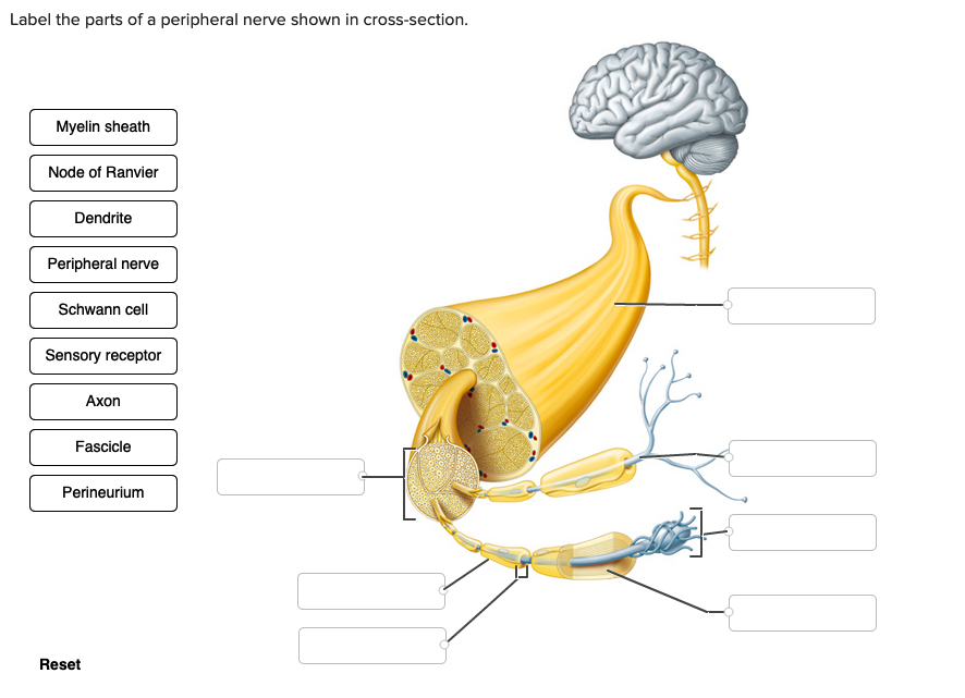
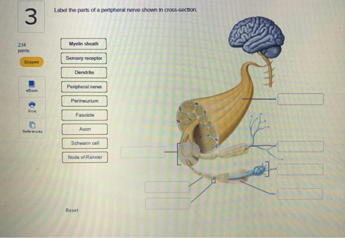
Label the Parts of a Peripheral Nerve Shown in Cross-section.
Solution for Draw and label a cross section of the spinal cord withits dorsal and ventral nerve roots. Start studying Peripheral nerve slide- Cross-Section.
Solved Label The Parts Of A Peripheral Nerve Shown In Chegg Com
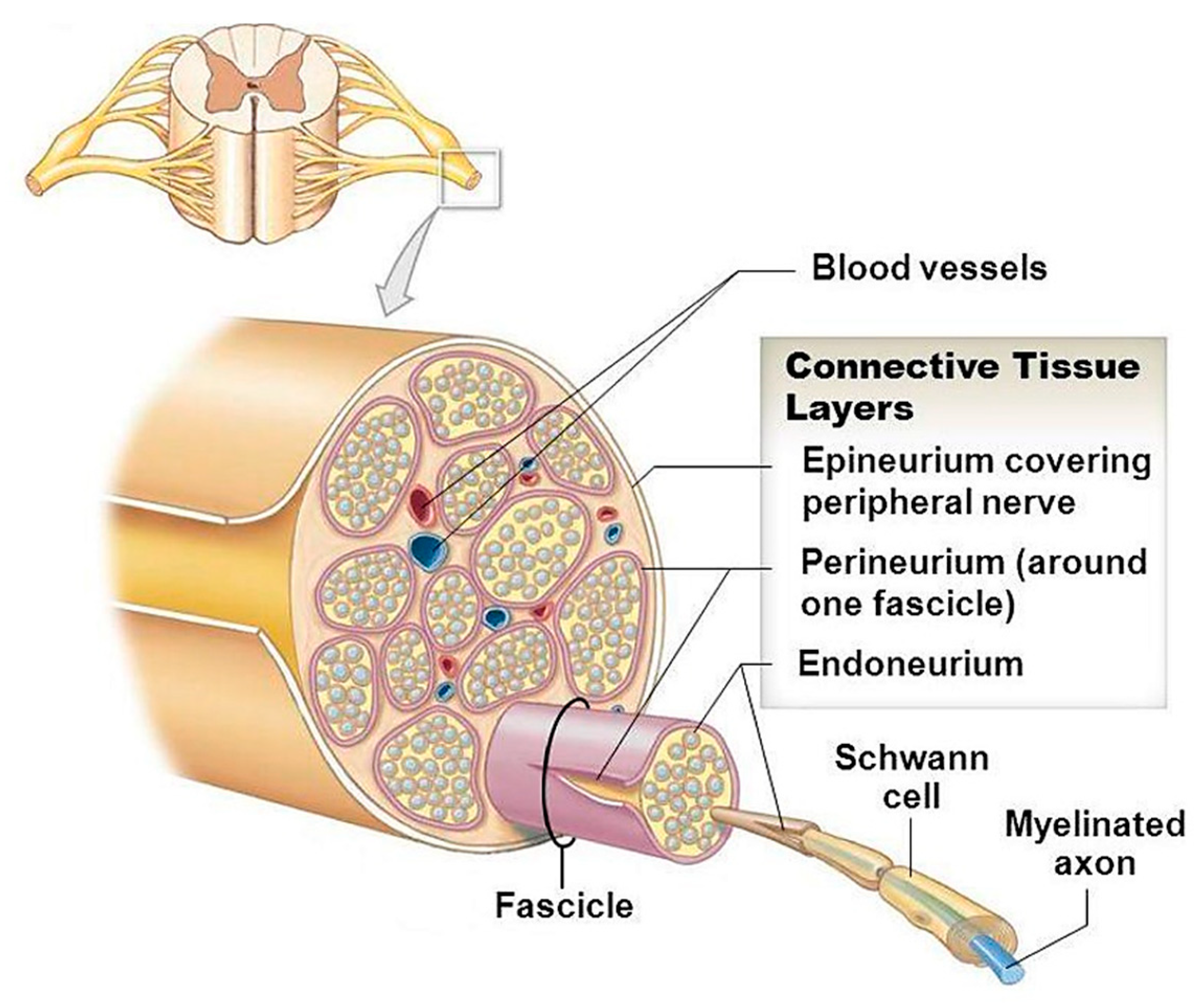
A whole nerve is surrounded by connective tissue called the epineurium not shown by this slide.
. The latter of which can either be sympathetic fight or freight response or. The spinal cord which consists of the major nerve tract of vertebrates runs down from the bottom of the brain through the passageway of the spinal column. Note the presence of neuronal fascicles each of which is a bundle of nerve fibers and note that the organ itself is a bundle of fascicles.
This is a peripheral nerve seen in cross-section. Diagram of a peripheral fiber showing a longitudinal section of parts of two adjacent Schwann cells and the axon they ensheath. Label the parts of a peripheral nerve shown in cross-section.
The dense pink-colored band is the perineurium. Label the parts of a peripheral nerve shown in cross-section. Start your trial now.
In the transverse section find epineurium perineurium and endoneurium see Figs. Can be inflamed in autoimmune rheumatologic disorders. Learn cross section nerve with free interactive flashcards.
Inner layer of posterior wall of eye see Retina in Cross Section contains receptors that convert light energy into signals that brain can interpret. On glass slide 51 in your Histology slide box longitudinal and transverse sections of a sciatic nerve from a horse are evident triple stain. Peripheral nerve monkey ls HE.
Inferior hypogastric plexus 214 points Cranial nerve VII Celiac plexus BOD Cranial nerve III Cranial nerve X Vagus Pelvic nerves. Wavy tissue poorly stained because of the high lipid content of the myelin axons surrounded by myelin sheaths. Label the parts of a peripheral nerve shown in cross section Myelin sheath Sensory receptor Dendrite Peripheral nerve Perineurium Fascicle Axon Schwann cell Node of Ranvier Label the features of the parasympathetic pathways.
In the peripheral nervous system axons are bundled together in structures called nerves. For each cranial nerve place the label in the appropriate box categorizing it based on function. This schematic depicts hypothesized routes nodes of.
Vascular layer that nourishes outer retina. This area is made up of all the nerve fibers that direct the reflex actions and convey the impulses that go back and forth to the brain. Unmyelinated nerve fibers which typically occupy at least 5 of a nerves cross-section can be visualized as axons without surrounding myelin sheaths.
Cranial nerves innervate parts of the head and connect directly to the brain. Schwann cell myelin sheath Nucleus of Schwann cell. Peripheral nerves consist of bundles of myelinated and non-myelinated nerve fibers enveloped by connective tissue.
At this magnification one can appreciate the general organization of the organ. Myelinated Nerve with Endoneurium and Perineurium cross section. A layer of connective tissue called the perineurium pn surrounds each fascicle.
Choose from 5000 different sets of cross section nerve flashcards on Quizlet. Learn vocabulary terms and more with flashcards games and other study tools. First week only 499.
Cranial nerves are typically assigned Roman numerals from 0 to 12. The nerves attached to the brain are the cranial nerves which are primarily responsible for the sensory and motor functions of the head and neck one of these nerves targets organs in the thoracic and abdominal cavities as part of the parasympathetic nervous system. Peripheral nerve fibers are grouped based on the diameter signal conduction velocity and myelination state of the axons.
Select View 50 um T. Dendrite Peripheral nerve Perineurium Axon Fascicle Sensory receptor Schwann cell -. The corpora quadrigemina cerebral peduncles and red nucleus are parts of the __ of the brainstem.
Superior gluteal nerve inferior gluteal nerve sciatic nerve posterior femoral cutaneous nerve pudenal nerve. Diameter Conduction Velocity Myelination State. Label the nerves of the lumbosacral plexus.
This is a Peripheral Nerve cross-section please use ARROWS to label the following. Spinal Cord Cross Section. Label the nerves of the brachial plexus.
The 40X image shows a cross section through four fascicles f that are part of a nerve. Between the axons you will see delicate connective tissue and an occasional fibroblast which constitute the endoneurium. Midbrain _ crevical nerves _ thoracic nerves _ lumbar nerve.
Scan the spinal cord. Peripheral nerve monkey cs HE. At the periphery of the fascicle observe the perineurium made up of several layers of flattened cells.
This is peripheral nerve. The central nervous system is comprised of the brain and spinal cord while the peripheral nervous system includes all spinal and cranial nerve fibres providing end organ innervation. Of note the peripheral nerve is also further subdivided into the somatic and autonomic divisions.
It is a highly specialized layer that acts as a barrier and protects the nerve from the environment. Draw and label a cross section of the spinal cord withits dorsal and ventral nerve roots. Iliohypogastric nerve ilioinguinal nerve genitofemoral nerve lateral cutaneous nerve of thich femoral nerve.
Collagenous outer layer of wall of eye.
Solved Label The Parts Of A Peripheral Nerve Shown In Chegg Com
Applied Sciences Free Full Text Bioactive Glasses And Glass Polymer Composites For Neuroregeneration Should We Be Hopeful Html

The Peripheral Nervous System Anatomy And Physiology


Comments
Post a Comment